Pages

Osteosarcoma

Before tumor removal and reconstructive surgery:


After tumor removal and reconstructive surgery:


There are some humans that don't get this good of a nose job.

Manhattan Cat Specialists
reade more... Résuméabuiyad

Manhattan Cat Specialists Fall 2010 e-Newsletter

UPDATE:  We have sent out our Manhattan Cat Specialists e-Newsletter - Fall 2010.  To view it, click here: http://conta.cc/cIw5eF

If you would like future newsletters (four a year), please add your email into the box below and press GO.


Email Newsletter icon, E-mail Newsletter icon, Email List icon, E-mail List icon
Sign up for our Manhattan Cat Specialists E-Newsletter


reade more... Résuméabuiyad

The Feline Patient - A Little Light Reading

Hot off the presses, the Fourth Edition of the ever-popular "The Feline Patient" arrived at my veterinary hospital today.  The book has a whopping 330 chapters.  Not surprisingly, my favorite chapter was chapter 149.


It is a fascinating read, and at 7 pounds (literally!), it makes an excellent door-stop.

reade more... Résuméabuiyad

Dance of the Kitty Cat's Ear Mites

We got a great microscopic image today of an insane swarm of ear mites


Ear mites are pesky, microscopic bugs that can live in your kitten’s ears, causing itching and relentless discomfort. Severe infestations can cause rupture of the eardrum and inflammation of the middle ear, resulting in balance and coordination problems. Often manifesting as an accumulation of dry, brown, crusty material in the ear canals, ear mites can cause kittens to scratch their ears so vigorously as to cause bleeding. Fortunately, they are easily diagnosed by your veterinarian, and have become easier to treat due to the recent development of new topical (on the skin) and otic (in the ear) medications.
reade more... Résuméabuiyad

Mack the Knife (Part 2)

( Warning: graphic medical images included in this post, please be advised. )

In my last blog post, I started telling the tale of Mack, the cat with malignant, metastatic mast cell tumor. Amazingly, Mack’s badly affected foot had healed after surgery,


however, a new tumor had popped up beneath his right eye.


This was worrisome because the tumor had the potential to grow beyond the lid and irritate the cornea. This tumor had to be removed while it was still small enough. The location – right below the eye – was difficult, because once the mass was removed, the defect left in the skin, when closed, could cause the lower lid to become distorted and roll outward.


As usual, Mack was a handful, the recent increase in handling having no effect on his feral nature. We gently anesthetized him, and I very meticulously removed the mass on his eyelid, making sure not to go too far to the edge. When I was finished, it looked pretty good!.

The lower lid was rolled out a little, but when he woke up and started blinking his eye, the lid became more and more natural looking, and you could barely tell anything had been done.

Unfortunately, Mack developed several more tumors on his skin. Chemotherapy options are limited for this type of tumor, and in a cat with such a feral temperament, medicating him regularly would be nearly impossible. Once he’s out and about in the apartment, you never see him. Forget about giving him pills or liquid. In discussing this with a veterinary oncologist, the only practical option would be to give him a long-acting steroid injection every 3 weeks, to see if it would have any effect on the tumors. We’re just about to begin this course of treatment. In a future blog post, I will give you an update on his condition.
reade more... Résuméabuiyad

Mack the Knife (Part 1)

( Warning: graphic medical images included in this post, please be advised. )

One of my favorite clients is an elderly woman we’ll call Mrs. B. She’s been coming to my practice for years. She started out with 20 cats. Slowly over time, her menagerie has gotten smaller and smaller, with cancer being the main culprit.

One of her remaining cats is a scraggly old male named Mack. Mack was taken off the street as a feral cat 17 years ago. You’d think, after 17 years, the cat would realize that Mrs. B was a friend and not a foe, but no, he hasn’t. Mrs. B hasn’t been able to touch this cat for 17 years. She never sees him. Occasionally, if she comes home and turns on the light fast enough, she’ll catch a glimpse of him as he scurries away like a cockroach. The only time she gets to touch him is when she brings him to see me. For that, she has to enlist the aid of a friend (who happens to have once been a veterinary technician). Trapping him and pushing him into a cat carrier is an ordeal, however, at 17 years of age, he’s lost much of his fighting spirit and he eventually yields to his pursuers.

Recently, on one of her rare glimpses of Mack, she noticed that he was limping on one of his front legs. Despite being three-legged lame, Mack put up a fuss about being corralled. When she finally brought him into me, it was clear that something was terribly wrong with Mack’s left front foot. A giant, unusual looking tumor.

 
As you can see in the photo, there’s a big pink tumor in the space between the main pad and toes. Further up the foot, there’s another pink fleshy tumor, surrounding the dewclaw. Interestingly, he had a similar looking fleshy mass in the middle of his back. 


I recommended that we remove and biopsy the mass on the back, and try to remove (and biopsy) the mass on the foot. Removing the mass on the back would be easy. There is a lot of skin in that area, and pulling the skin defect together with a few stitches would be no problem. The foot would be more of a challenge.

There’s no extra skin on the foot, so removal of skin in the foot area often leads to a defect that can be difficult to close. In Mack’s case, we didn’t have much of a choice. If unaddressed, this mass would continue to grow and become infected.

Removing the mass around the dewclaw wasn’t that bad. Surprisingly, I was able to pull the skin and close the defect without much tension on the skin. The foot paw itself was another matter. The mass was pretty infiltrative, and by the time I managed to get it all out, I was left with a large defect that would be a challenge to close.

The only way I could conceivably close this was to cut the webbing between toe #1 and toe #2, and then wrap the skin around each toe and suture it closed.


I was then able to wrap the remaining piece of skin around both toes #3 and 4, fusing them together. We then bandaged the foot and kept Mack hospitalized for a few days in our facility, so that we could change the bandage regularly and watch the foot.

Unfortunately, the biopsy came back as a mast cell tumor. These are mysterious tumors that occasionally arise in feline skin and are almost always benign. Removal is recommended, because they do have the theoretical potential to spread to internal organs like the spleen and the gastrointestinal tract. Mack’s mast cell tumor was different, though. His was a malignant mast cell tumor, and it had already spread to other parts of his body. In fact, the piece of tissue from his foot was identical, under the microscope, to the piece from the middle of the back. They were the same tumor, in different parts of the body, indicating metastasis (i.e. spreading).

Although euthanasia would be justified, especially in a case like Mack’s, where chemotherapy and regular checkups and monitoring wouldn’t be possible due to his feral nature, Mrs. B agreed to take Mack home, keep him in a cage so that he wouldn’t disappear for weeks at a time, and bring him back for us to check on his foot. I fully expected the foot to look terrible at the next visit, because I could not remove the entire tumor, and I expected it to grow back. To my surprise, the foot looked great!


The sutured toes were healing. Only the middle toe hadn’t healed.


I didn’t think it would, because it looked like the open area of the toe was infiltrated with tumor tissue, but at the next visit, you can see that it healed even further.


But, more trouble was in store for Mack. Stay tuned.
reade more... Résuméabuiyad

Cleft Palate - It's either there, or it’s not. (It was)

Let me tell you about an interesting (and strange) case I had a few weeks ago. A young couple (actually, they weren’t all that young, but now that I’m 50, everyone seems young.)( Sigh.) brought their 7 month old kitten in for a second opinion. Their little kitty had a snotty nose and had been sneezing for weeks. The nasal discharge was primarily from the right nostril. The previous veterinarian prescribed antibiotics, which helped a little, but never cleared it up completely. So he tried a second course of antibiotics, which again caused a little improvement, but it went right back to the profuse sneezing and nasal discharge as soon as the antibiotics were through. So he tried a different antibiotic. The owners were getting frustrated at this approach (you can’t really blame them), so they brought the cat to my hospital, Manhattan Cat Specialists.

I examined the kitty, and found her to be a bright, energetic, healthy little thing. Except for the snotty right nostril. While standing on the exam table, she sneezed and sprayed our wall with the nasal discharge. Lovely.

Young cats often develop polyps in their nasal cavity, and these lead to chronic snotty nose and noisy breathing. I was thinking that this might be the case with this kitten, but listening to her breathe, it didn’t sound like a polyp. Cats with polyps breathe like bulldogs or pugs, i.e. very noisy. You can hear them across the room. This kitty was breathing normally. On my examination, however, I found the problem right away: in the roof of her mouth was a tiny little hole! This is known as a cleft palate.

A cleft palate is a congenital defect; kittens are born with it. If the hole is big, food that is taken into the mouth will go through that hole into the nasal cavity, irritating the sinuses and causing infection, which is what was happening here. I pointed out the hole to the clients. There was much oohing and aahing.

Treatment requires surgical repair. This is something that I have only attempted once, 20 years ago, and I didn’t feel comfortable repairing this one myself. I recommended a well-known referral center (whose name I will withhold). This center has a department specifically for oral/dental problems in cats.

A few days later, I received a faxed report from the doctor that examined the kitten. To my great surprise, they said that they anesthetized the cat, examined the mouth thoroughly, probed every tooth, and concluded that there was no cleft palate! Since this cat did not have a surgical disease, they concluded, they did not do any surgery, and they were going to send her back to me for further investigation of the cause of her respiratory problems.

Okay, this is weird. This is not some subjective interpretation of an ultrasound image, where two doctors differ in opinion. This is a hole. Either it’s there or it’s not. So I called and spoke to the doctor. He happened to be an intern. He told me in person (well, on the phone) that he looked and there simply wasn’t a cleft.

Silence.

I can hear him breathing on the phone. I know what he’s thinking. He’s thinking I’m crazy. Meanwhile, I know what I’m thinking. I’m thinking he’s crazy.

I call the owner. He said he thought it was weird, because he saw the hole with his own eyes during our exam. We thought, hmmm, it was so small. Maybe it closed up? But the cat still had the snotty nose and the sneezing. In the end, we decided to schedule the cat for rhinoscopy, a procedure where you look up the nostril using a rhinoscope – a long rigid tube with a light source and camera on the end.

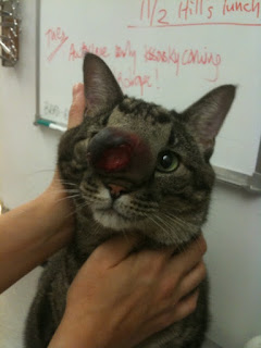
The cat comes in the following week. We anesthetize her and start preparing her for the rhinoscopy. As we open her mouth to insert the tracheal tube, what do I see on the roof of the mouth? The freakin’ cleft! Grrrrrr!!!


Fortunately, I had my trusty iPhone with me, and took a picture of the cleft, as you can see. I also took a picture with a wire inserted into it, so there is no mistaking it.


To be complete, we continue with the rhinoscopy, just to rule out a foreign body or tumor (highly unlikely) as the cause of the cat’s nasal problem.

I am a strong proponent of the rule that “thou shalt not speak ill of another veterinarian”, there really is no way around this one. Again, this is not a case of differing subjective opinions. It’s like pregnancy: either you are, or you’re not. This cat has a cleft. So I contacted the head of this referral institute by e-mail and by voice mail. My e-mail was very direct. Photos were attached.

I received a response instantly. The head of the institute was clearly embarrassed and contrite. He agreed to see the cat again, and to set things right. The problem now, though, is that cleft palates are rare in cats, and no one in their dental department had ever really done one. But…they were going to ask the surgeons in their surgery department if they wanted to tackle it. (They tried to convince me to recommend to the client a CT scan of the head to get a really good look inside the nasal cavity, because they had a super high-powered state-of-the-art top-of-the-line new CT scanner. I told him to knock it off; the cat’s problem is the cleft, so just fix it, dammit. He sheepishly backed down.)

The following day, I heard from one of their surgeons. She said that she hadn’t done one, but she had been reading about them and that she saw the photos, saw that it was a small one, and was up to the challenge. They agreed to do this at a major discount. I give them credit for that.

Well, this story has a happy ending. The surgeon successfully repaired the cleft, as you can see.

 
There’s a nice little row of dissolvable sutures in the palate. The cat’s nasal discharge and sneezing immediately resolved. The owners said that the cat is not only no longer sneezing and slinging snot everywhere, but she is just happier in general; a totally different cat.

I love when cases end like this.
reade more... Résuméabuiyad